

Magnetic Resonance Imaging (MRI) is an imaging technique that is based on the principles of magnetic resonance. In medical diagnostics it is used to obtain images of organs inside the human body, similar to X-ray tomography. Unlike the latter, in which the images reflect the electron density, MRI gives an image of the proton density. By a suitable choice of experimental parameters, MRI provides the ability to create additional contrast in the images between different chemical and physical environments of the protons. This feature makes MRI of interest for spatially resolved structure studies of various materials such as Polymers. For obtaining NMR images, it is necessary to superimpose the static magnetic field of the NMR spectrometer with pulsed magnetic field gradients, causing the resonant frequencies of the protons to be a function of their position within the field gradient. Since the gradients of our equipment are significantly stronger than those used in medical imaging, we achieve resolutions in the order of 10 μm, whereas typical resolutions in medical imaging are in the order of 1 mm.
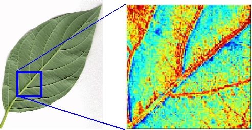
Dogwood-Leaf: Photo and MRI-image showing iron concentration